HIP JOINT DISEASES
Femoroacetabular Impingement
It occurs due to abnormal contact and relationship between the femoral neck and the edge of the acetabulum. And it is painful condition. It is the most common cause of hip pain especially in young individuals who are involved in sports (ballet, martial arts, gymnastics, etc.) where the hip is exposed to excessive movements. It is common for the labrum to rupture with this excessive contact, which increases the depth of the acetabulum. When the labrum tear occurs, joint cartilage damage also occurs. Thus, osteoarthritis occurs in the hip joint.
In radiological appearence; Measurements are taken to detect hump or acetabulum retroversion at the head and neck junction. According to the MRI results, the labrum tear and cartilage damage are evaluated. In treatment; NSAID drugs and activity restriction should be applied to relieve edema and pain that occurs first in the hip. Surgical treatment is applied to patients who do not benefit from this treatment. The aim of surgical treatment is to shave the shape formed in the femoral head and neck junction and to repair the labrum tear in the acetabulum. If this labrum tear cannot be repaired, it should be removed. These surgical procedures can be performed with closed arthroscopic method or with open controlled hip dislocation.
Developmental Dysplasia Of The Hip (Hip Dislocation)
It is the name given to the condition that the femoral head is not adequately covered by the acetabulum. With the deterioration of joint harmony; while the patient had been walking, the amount of load on the femoral head cartilage changes. Thus a more than normal load is placed on a small area. In the evaluation of hip dysplasia, the degree of dislocation on ultrasound and radiographs and the amount of coverage of the femoral head are important. Among the treatments applied in infancy and the treatments in adulthood are different. Therefore, it will be crucial to detect and treat in this disease in early infancy. Infant hip dysplasia may be treated with only the Pavlik bandage in many patients. Hip replacement surgery is required while adulthood. In young adulthood, periacetabular osteotomies such as Ganz, Tönnis or Bernese are applied to for increase the coverage of the acetabulum. With this method, advanced osteoarthritis of the hip is prevented.
Osteonecrosis Of The Femural Head
It is a disease that usually occurs in people younger than 50 years of age and is caused by the deterioration of blood supply to the femoral head due to various reasons. As a result, the cartilage of the femoral head softens and becomes deformed. Therefor thus, it causes rapid osteoarthritis of the hip joint. This is the second most common reason for hip replacement. There are many conditions that cause this disease (trauma, alcohol, cortisone, hit, sickle cell anemia, etc.). But in many patients, no cause is found. MRI is important for diagnosis in the early stages and in follow-up. However, radiography changes occur in advanced stages.
The stage of the disease is vital for the treatment. In the early stages, non-prosthetic treatments such as core-decompression, vascular fibula application or hip osteotomies can be applied. After the collapse of the femoral head, total hip replacement is the only treatment option.
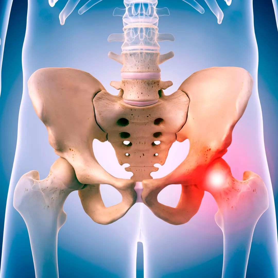
Hip Osteoarthritis (Coxarthrosis)
Of the hip joint; It is a painful condition that occurs in the groin and hip due to the deterioration of the cartilage structure due to various reasons. Generally patients says the pain feels like been walking a long distance. They indicate that have difficulty while wearing shoes and socks. And sometimes they feel the hip pain in the knee. Due to the loss of cartilage due to arthrosis, leg shortening and limping may be felt on the same side. On examination, hip movements, especially internal rotation, are limited and painful.
Classical coxarthrosis findings on radiographs can be listed as follows; joint space narrowing, subchondral sclerosis, osteophyte at the joint margins, cyst formation and deformation of the femoral head and/or acetabulum.
The age of the patient and the degree of calcification are important when deciding on the treatment. According to the stage of the disease, treatment is started with weight loss pain reliever/edema-solving drugs. If there are activity modifications, shortness compensation and hip adductor stretching, abductor strengthening exercises are started. If the patient does not benefit from these treatments and the quality of life deteriorates due to hip pain, surgical treatment options are recommended to the patient. Non-prosthetic hip-sparing osteotomies should be considered in surgical treatment, especially in young and active patients. For example, in a young patient with acetabular cover insufficiency because of hip dislocation; It reduces the load on the femural head and cause hip calcification. In patients with advanced age; with hip arthrosis who do not benefit from weight loss, drug therapy and physical therapy, the most appropriate treatment option is total hip replacement.